Skip to content
Division of Health AIDivision of Health AI
TeamResearchPublicationsJoin
Theme
Division of Health AIDivision of Health AI

Site

  • Team
  • Research
  • Publications
  • Join

Affiliated institutions

  • Feinstein Institutes (opens in new tab)
  • Northwell Health (opens in new tab)
  • Zucker School of Medicine (opens in new tab)

© 2026 Division of Health AI, Northwell Health. All rights reserved.

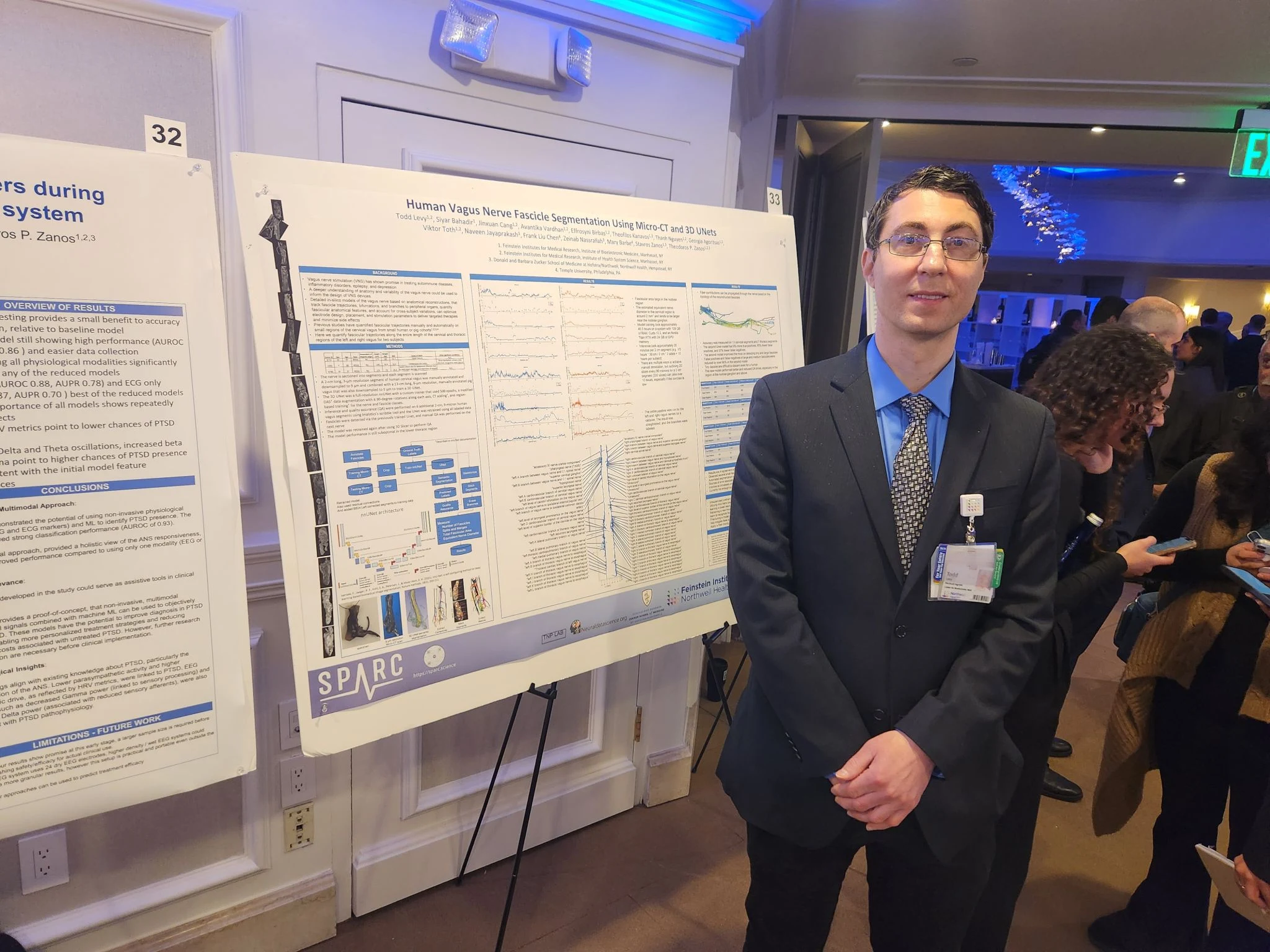
Todd Levy beside his poster on human vagus nerve fascicle segmentation at a SPARC meeting

Publications

Human vagus nerve fascicle segmentation, SPARC · Photo: Division of Health AI

2 publications · filtered

Clear filters
Brain Stimulation · Feb 2023

Organ- and function-specific anatomical organization of vagal fibers supports fascicular vagus nerve stimulation

(opens in new tab)

Background: Vagal nerve fibers traveling in distinct fascicles innervate different organs and regulate specific functions. Current vagus nerve stimulation (VNS) therapies activate vagal fibers non-selectively, often resulting in reduced efficacy and side effects. Objective: To characterize the anatomical organization of vagal fibers and demonstrate fascicle-selective VNS. Methods: We used quantified immunohistochemistry, micro-computed tomography imaging, and multi-contact cuff electrodes to map fascicular organization and perform selective stimulation in swine. Results: Myelinated afferents and efferents occupy separate fascicles. Larynx-, heart-, and lung-specific fascicles are spatially separated and progressively merge. Fascicle-selective VNS elicited organ-specific physiological responses with radially asymmetric compound action potentials. Conclusions: Fascicular VNS enables selective modulation of specific organs and functions, offering improved efficacy and reduced off-target effects compared to conventional non-selective VNS.

Brain Stimulation

Feb 2023
Annual International Conference of the IEEE Engineering in Medicine and Biology Society (EMBC) · Jul 2020

Single-axon level automatic segmentation and feature extraction from immunohistochemical images of peripheral nerves

(opens in new tab)

Quantitative descriptions of the morphology and structure of peripheral nerves is central in the development of bioelectronic devices interfacing nerves. While histological procedures and microscopy techniques yield high-resolution detailed images of individual axons, automated methods to extract relevant information at the single-axon level are not widely available. A segmentation algorithm was implemented that allows for feature extraction in immunohistochemistry (IHC) images of peripheral nerves at the single fiber scale. Features extracted include short and long cross-sectional diameters, area, perimeter, thickness of surrounding myelin and polar coordinates of single axons within a nerve or nerve fascicle. The algorithm was evaluated using manually annotated IHC images of 27 fascicles of the swine cervical vagus; the accuracy of single-axon detection was 82%, and the classification accuracy of fiber myelination was 89%.

Annual International Conference of the IEEE Engineering in Medicine and Biology Society (EMBC)

Jul 2020

More

Explore the researchMeet the teamInternships and PhD training