Division of Health AINorthwell Health
AboutTeamResearchPublicationsJoin Us
Division of Health AI

Northwell Health

  • About
  • Team
  • Research
  • Publications
  • Join Us
  • Feinstein Institutes
  • Northwell Health

© 2026 Division of Health AI, Northwell Health

Admin

Division ofHealth AI

Northwell Health

Feinstein Institutes for Medical Research | Institute of Health System Science | Institute of Bioelectronic Medicine Artificial Intelligence and Machine learning for early diagnosis, clinical outcomes prediction, and personalized therapies

ResearchTeamPublicationsJoin us

Research

View all

Point-of-care AI

Develop clinical decision support tools to diagnose and predict clinical outcomes and trajectories. This vertical focuses on preventing in-hospital deterioration through AI-driven monitoring, including a deep-learning model to reduce unnecessary overnight vitals checks, a deterioration prediction model trained on over 1.5 million hospitalizations that outperforms Epic's Deterioration Index by 25%, and a wearable-based continuous monitoring system funded by a $3.2M NIH grant that predicts clinical alerts 17 hours in advance with over 94% sensitivity.

Operational AI

Develop operational decision support tools to provide enterprise insights and assist in health system wide issues. This vertical addresses the nursing workforce crisis through AI-driven forecasting, using DeepAR models trained on 5 years of historical data to predict nursing demand and attrition up to 12 months ahead. The preemptive hiring strategy is estimated to save close to $10 million annually across just 10 of Northwell's 400+ units by reducing reliance on flex staff and overtime. Also includes unsupervised clustering to phenotype nurse attrition and identify modifiable factors.

Preclinical AI

Develop algorithms that use neural recordings from preclinical models to diagnose and predict disease states. This vertical focuses on decoding vagus nerve signals related to inflammation and metabolic states, building on foundational work published in PNAS showing that cytokine-specific neural activity can be decoded from the cervical vagus nerve. Includes chronic vagus nerve recording in implanted mouse models and predicting disease severity and treatment efficacy for conditions such as rheumatoid arthritis using vagus nerve stimulation.

Autonomic Nervous System AI

Develop algorithms that use non-invasive physiological data to diagnose disease presence and severity and predict treatment efficacy. Key projects include predicting vagus nerve stimulation treatment response for drug-resistant epilepsy patients, and using machine learning on autonomic nervous system biomarkers to detect and quantify PTSD presence and severity from physiological signatures. This work uses heart rate variability and other non-invasive measures to build parsimonious diagnostic and prognostic models.

Anatomical Data AI

Develop accurate in-silico models of human anatomy using multimodal imaging and AI. This vertical is creating the most detailed human vagus nerve digital twin in existence, processing over 4 million microscopy and micro-CT images (approximately 200 terabytes of data) from 58 vagus nerves (left and right from 29 cadavers) to map the spatial arrangement of fascicles and fibers from the cervical and thoracic trunks to organ-level branches. Funded by a $6.7M NIH SPARC REVA grant, this work leverages deep learning architectures—including nnU-Net for 3D micro-CT segmentation and Mask2Former for individual identification of myelinated and unmyelinated axonal cross sections in immunohistochemistry data—to quantify vagus nerve anatomy and inform the design of next-generation selective vagus nerve stimulation devices.

Paper Highlight

Nature Communications

Beyond episodic early warning systems: a continuous clinical alert system for early detection of in-hospital deterioration

A wearable-based deep learning model that identifies the onset of clinical deterioration earlier than traditional early warning systems, predicting adverse outcomes up to 17 hours in advance with over 81% accuracy.

Selected Publications

View all
Nature Communications

Control of spatiotemporal activation of organ-specific fibers in the swine vagus nerve by intermittent interferential current stimulation

Vagus nerve stimulation (VNS) is emerging as potential treatment for several chronic diseases. However, limited control of fiber activation, e.g., to promote desired effects over side effects, restricts clinical translation. Towards that goal, we describe a VNS method consisting of intermittent, interferential sinusoidal current stimulation (i2CS) through multi-contact epineural cuffs. In experiments in anesthetized swine, i2CS elicits nerve potentials and organ responses, from lungs and laryngeal muscles, that are distinct from equivalent non-interferential sinusoidal stimulation. Resection and micro-CT imaging of a previously stimulated nerve, to resolve anatomical trajectories of nerve fascicles, demonstrate that i2CS responses are explained by activation of organ-specific fascicles rather than the entire nerve. Physiological responses in swine and activity of single fibers in anatomically realistic, physiologically validated biophysical vagus nerve models indicate that i2CS reduces fiber activation at the interference focus. Experimental and modeling results demonstrate that current steering and beat and repetition frequencies predictably shape the spatiotemporal pattern of fiber activation, allowing tunable and precise control of nerve and organ responses. When compared to equivalent sinusoidal stimulation in the same animals, i2CS produces reduced levels of a side-effect by larger laryngeal fibers, while attaining similar levels of a desired effect by smaller bronchopulmonary fibers.

Translational Vision Science & Technology

Artificial Intelligence-Driven Differentiation Between Uveal Melanoma and Nevus Based on Fundus Photographs: A Systematic Review and Meta-Analysis

Uveal melanoma (UM) is the most common intraocular malignancy in adults, with high metastatic risk and poor prognosis. Current screening and triaging methods for melanocytic choroidal tumors face inherent limitations, particularly in regions with limited access to specialized ocular oncologists. This systematic review and meta-analysis evaluated artificial intelligence-driven approaches for differentiating uveal melanoma from nevus based on fundus photographs. Analysis included machine learning models with pooled sensitivity of 85% (95% CI 82–87%), specificity of 86% (82–88%), and a C-index of 0.87 (0.84–0.90), with convolutional neural networks as the main method used. Deep learning models achieved AUC scores of 94-95%, outperforming ophthalmologists using standard risk assessment criteria.

Interested in joining or collaborating? Get in touch

Nature Communications

Beyond episodic early warning systems: a continuous clinical alert system for early detection of in-hospital deterioration

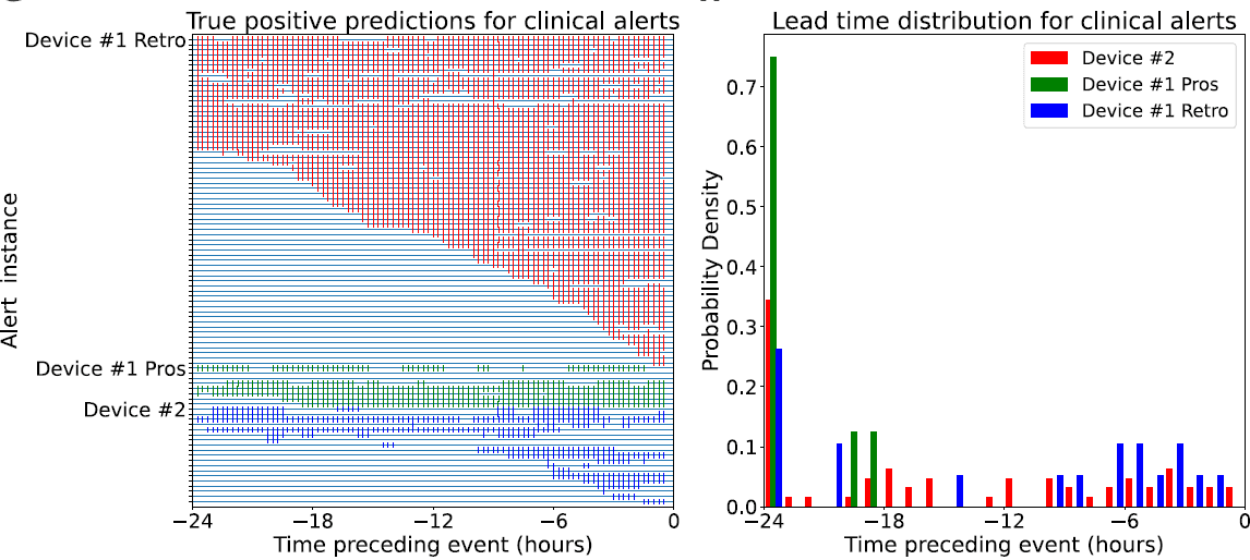
Efficient patient monitoring on medical-surgical wards is crucial to prevent adverse events. Standard episodic inpatient assessment of vital signs can miss changes in health status and delay risk recognition. This study developed a wearable-based deep learning model using only 9 inputs to identify the onset of deterioration earlier than traditional early warning systems. The model could generalize to produce clinical alerts ahead of rapid response team (RRT) interventions, unplanned intensive care unit (ICU) transfers, intubations, cardiac arrests, and in-hospital deaths. Using multiple stages of validation on 888 adult non-ICU inpatient visits, the RNN model predicted both periods of elevated MEWS scores (ROC AUC 0.89 +/- 0.3, PR AUC 0.58 +/- 0.14) and adverse clinical outcomes (accuracy: 81.8%) up to an average of 17 hours in advance.

International Journal of Neural Systems

Longitudinal characterization of compound action potentials in chronic vagus nerve recordings in mice

Even though extensively documented in acute experiments, ongoing vagal activity has not been characterized longitudinally over days or weeks in mice, a preferred preclinical model. This study presents a chronic recording model to record compound action potentials (CAPs) from the mouse vagus nerve for up to 6 months in both anesthetized and awake animals, with stable signal-to-noise ratios and half-rise times. The approach allows for longitudinal analysis while tracking individual CAPs across multiple days, their firing rates and phase-locking characteristics with other physiological signals, and in the awake case, movement using unsupervised machine learning models. Results reveal diverse CAP populations with varying degrees of physiological coupling, providing a valuable platform to investigate how vagal activity may be modified based on disease severity and develop closed-loop VNS by predicting flare-ups and tracking stimulation efficacy.